Asbestos Toxicity
Clinical Assessment - Tests
Course: WB 2344
CE Original Date: January 29, 2014
CE Renewal Date: January 29, 2016
CE Expiration Date: January 29, 2018
Download Printer-Friendly version [PDF - 1.28 MB]
| Previous Section | Next Section |
Learning Objectives |
Upon completion of this section, you will be able to
|
|||||||||||||
Introduction |
The most important tests in diagnosing asbestos-associated disease are
Other tests and procedures that are sometimes used to diagnose asbestos-associated diseases by specialists in cases that require further work-up include
|
|||||||||||||
Screening Pulmonary Function Tests |
Screening pulmonary function tests are useful for finding restrictive deficits most commonly associated with asbestosis (see table). Findings may include a reduction in forced vital capacity (FVC) with a normal forced expiratory volume (FEV) in one second FEV1/FVC ratio. Some sources report abnormal pulmonary function tests in 50% to 60% of patients with asbestosis [Ross 2003]. In some cases, combined patterns of restrictive and obstructive disease may be seen. For further assessment of whether a patient has a restrictive abnormality and asbestosis, additional, more specialized tests may be required.
Consider consulting a pulmonologist if the diagnosis is unclear, if there is a rapid decline in pulmonary function, or if there is a potential need for a tissue biopsy or BAL, such as in cases where lung cancer, mesothelioma, or an infection is suspected. The pulmonologist may recommend more extensive pulmonary function tests.
|
|||||||||||||
Chest Radiograph |
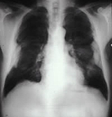
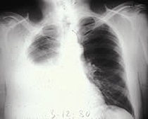
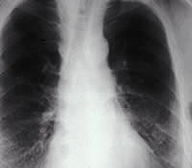
The chest radiograph is used primarily to find, localize, and assess the extent of structural changes associated with asbestos-caused chest diseases (asbestosis, non-malignant pleural disease, mesothelioma, and lung cancer). Diagnosis of asbestosis should mostly but not totally be based on radiographic findings, per the diagnostic criterion of the American Thoracic Society. In 10% to 15% of cases, an asbestos-associated pulmonary function abnormality can occur without definite radiologic change [Ross 2003]. The association of pleural thickening and calcification with interstitial changes enhances diagnostic accuracy of asbestosis. During the latter half of the past century, the International Labour Office (ILO) developed a system for radiographic classification of the pneumoconiosis. It now includes a set of standard digital radiographic images or a set of standard plain film images. [ILO 2011, NIOSH 2011b] Persons certified by the National Institute of Occupational Safety and Health (NIOSH) as proficient in the use of this rating system are called "B Readers." Most epidemiological studies of pneumoconiosis use B Readers to classify radiographs according to the ILO system. A current list of B Readers can be found at http://www.cdc.gov/niosh/topics/chestradiography/breader-list.html. Classification of asbestosis by chest radiography should be guided by the ILO system. A list of typical chest radiograph findings for each of the asbestos-associated diseases is in the table below.
|
|||||||||||||
CT and HRCT |
In some cases, computed tomography (CT) scans, including high-resolution computed tomography (HRCT) scans can facilitate diagnosis of asbestos-associated diseases such as lung fibrosis due to their greater sensitivity and specificity than conventional chest radiographs [Vierikko et al. 2010; Parris et al. 2009]. Because they are associated with higher doses of radiation than conventional chest radiographs and their cost-effectiveness and efficiency as screening tools have not been established, CT scans are not currently recommended in the United States for routine screening of asbestosis of the general population. They can be useful in further investigating abnormalities found on chest radiographs and in detecting abnormalities not seen on chest films of patients with dyspnea or pulmonary function abnormalities such as decreased DLco [Vierikko et al. 2010]. CT and HRCT scans are more sensitive and specific than chest radiographs. HRCT scans can be used particularly when abnormalities on a conventional chest radiograph are equivocal or when a conventional chest radiograph is normal in the face of unexplained lung function abnormalities in a patient with significant asbestos exposure [American Thoracic Society 2004]. They are especially useful in detecting
However, HRCTs are recommended for use in screening special populations at high risk of lung cancer such as people with smoking histories of 15 to 30 pack years and those with some occupational exposures such as asbestos, arsenic, chromium, silica, nickel, cadmium, berryllium and diesel fumes [Bach et al. 2012; NIOSH 2012b]. The utility of other imaging techniques such as ultrasound, gallium scanning, magnetic imaging, ventilation-perfusion studies, and positron emission tomography has not been established in asbestos-related disease. |
|||||||||||||
BAL and Lung Biopsy |
Bronchoalveolar lavage (BAL) is sometimes used by specialists to identify other possible causes for lung pathology. It can be used to assess exposure to asbestos by measuring the amount and type of asbestos bodies and asbestos fibers in the lavage fluid [American Thoracic Society 2004]. Not only is this a somewhat invasive procedure requiring fiberoptic bronchoscopy, but the laboratory procedures are not routinely available; special laboratory facilities and expertise are required. Lung biopsy is a definitive test used in the histopathological confirmation of asbestos-associated diseases. Lung biopsies are rarely used to diagnose asbestosis or pleural plaques, because diagnosis of these conditions is usually based on medical and exposure histories, findings from the physical examination, and other tests. Appropriate referral to a specialist is indicated if lung cancer or mesothelioma is suspected, since a lung biopsy will be indicated under these conditions. |
|||||||||||||
Blood Studies |
Blood studies may occasionally be useful for ruling out other causes of restrictive lung disease. |
|||||||||||||
Colon Cancer Screening |
Evidence increasingly shows that asbestos exposure increases an exposed patient's risk for colon cancer [IARC 2012]. Therefore, it is important to perform colon cancer screening on asbestos exposed patients starting at age 50 according to current guidelines [Agency for Health Care Research and Quality 2008]. The joint guidelines from the American Cancer Society, the U.S. multi-society task force for colorectal cancer, and the American College of Radiology can be found at: http://www.ncbi.nlm.nih.gov/pubmed/18322143 [Levin et al. 2008]. |
|||||||||||||
False Positives and False Negatives |
It is important to know what other conditions bear radiographic similarities to changes associated with asbestos-related disease (see table).
|
|||||||||||||
Attribution of Asbestos-Related Cause |
To help attribute pulmonary fibrosis to asbestos exposure, check the diagnostic guidelines suggested by the American Thoracic Society:
Bilateral calcified pleural plaques are usually attributable to asbestos exposure, but unilateral pleural plaques should motivate a search for other causes such as old tuberculosis, empyema, or hemothorax. CT scanning is useful to make a definite diagnosis of rounded atelectasis [Khan et al. 2013]. As stated previously, sets of diagnostic criteria like the Helsinki criteria can help in attributing an individual patient's lung cancer to asbestos exposure. Malignant mesotheliomas are, for all practical purposes, readily attributable to past asbestos exposure. |
|||||||||||||
Key Points |
|
|||||||||||||
Progress Check |
| Previous Section | Next Section |


