


Case #256 - July, 2009
 ShareCompartir
A 25-year-old refugee from Myanmar was screened for parasites at a local clinic in Tennessee. Stool specimens were collected in 10% formalin and polyvinyl alcohol (PVA) and sent to the state health laboratory for ova and parasite (O&P) work-up. Figures A-D show what was observed in a concentrated wet mount made from the formalin-preserved stool. Figures A and B show what was observed under brightfield microscopy; Figures C and D show the same fields, respectively, observed under UV microscopy. All images were captured at 400x magnification. The objects of interest measured on average 17 micrometers long by eight micrometers wide. What is your diagnosis? Based on what criteria?
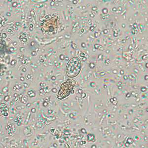
Figure A
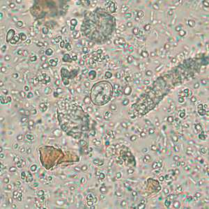
Figure B
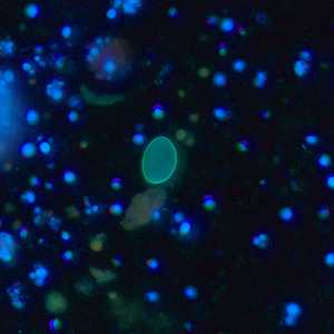
Figure C
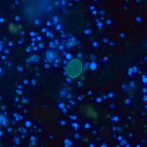
Figure D
Case Answer
This was a case of sarcocystosis caused by a member of the genus, Sarcocystis. The two species that cause intestinal sarcocystosis in humans are S. hominis and S. suihominis; the two species cannot be differentiated morphologically. Diagnostic features included:
- individual sporocysts within the size range for Sarcocystis spp. (15-20 micrometers long by 8-10 micrometers wide).
- the ability of sporocysts to autofluoresce under UV microscopy (Figures C and D).
Usually sporulated oocysts, each containing two sporocysts, are shed in the stool of the definitive host. These oocysts typically measure 15-20 micrometers long by 15-20 micrometers wide. Due to the fragile nature of the oocysts, often individual sporocysts are observed in stool specimens, as seen in this case.
More on: Sarcocystosis
Images presented in the monthly case studies are from specimens submitted for diagnosis or archiving. On rare occasions, clinical histories given may be partly fictitious.
DPDx is an education resource designed for health professionals and laboratory scientists. For an overview including prevention and control visit www.cdc.gov/parasites/.
- Page last reviewed: August 24, 2016
- Page last updated: August 24, 2016
- Content source:
- Global Health – Division of Parasitic Diseases and Malaria
- Notice: Linking to a non-federal site does not constitute an endorsement by HHS, CDC or any of its employees of the sponsors or the information and products presented on the site.
- Maintained By: