


Case #370 - April 2014
 ShareCompartir
A 21-year-old female presented with swelling of the eyelid, wrist, and knee. The symptoms started after returning from a three-month trip to Cameroon in 2011. A blood specimen was collected by her health care provider at 11:30 A.M. and smears were made and stained (NOS). Her eosinophilia at the time was 48%. A filarial IgG4 antibody test performed a month prior and was negative. Two worm-like structures were observed by laboratory technicians after scanning 10 stained thick smears. Images of both structures were captured (Figures A and B) and shared with CDC personnel. As follow-up to the images, a tube of EDTA blood was sent to the DPDx Team for further evaluation. Figure C shows what was found on a wet mount of the blood; Figures D and E are from thick and thin Giemsa-stained smears, respectively. What is your diagnosis? Based on what criteria?
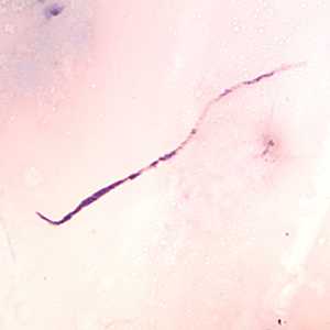
Figure A

Figure B
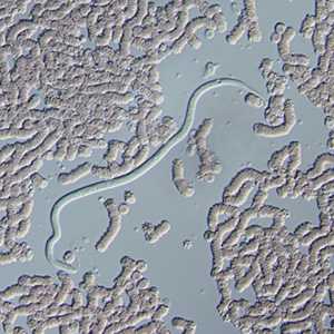
Figure C
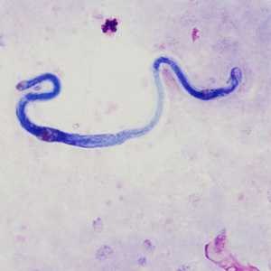
Figure D
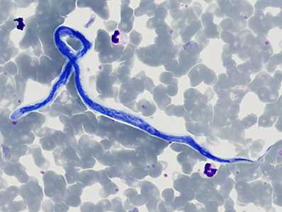
Figure E
Case Answer
This was a case of loiasis caused by Loa loa. Morphologic features shown included:
- presence of microfilariae in blood collected during the day.
- robust microfilariae with a short head space and a tapered tail with nuclei spaced irregularly to the tip of the tail (Figures A, B, D, and E)
- evidence of a sheath that did not stain with Giemsa (Figure C)
More on: Loaisis
This case and images (in part) were kindly provided by Purdue University, Indianapolis, IN.
Images presented in the monthly case studies are from specimens submitted for diagnosis or archiving. On rare occasions, clinical histories given may be partly fictitious.
DPDx is an education resource designed for health professionals and laboratory scientists. For an overview including prevention and control visit www.cdc.gov/parasites/.
- Page last reviewed: August 24, 2016
- Page last updated: August 24, 2016
- Content source:
- Global Health – Division of Parasitic Diseases and Malaria
- Notice: Linking to a non-federal site does not constitute an endorsement by HHS, CDC or any of its employees of the sponsors or the information and products presented on the site.
- Maintained By: