


Case #384 - November 2014
 ShareCompartir
A 73-year-old woman presented to a local clinic in Ghana with a skin nodule that had developed adjacent to the resection site of a previous low-grade malignant skin tumor. Biopsy specimens were collected and sent to a lab in the U.S. for histologic processing, including sectioning and staining with hematoxylin-and-eosin (H&E). Images of suspect structures were captured and sent to the DPDx Team for diagnostic assistance. Figures A-E show several of the images that were received for analysis. Neither sizes nor magnifications were provided with the images. What is your diagnosis? Based on what criteria?
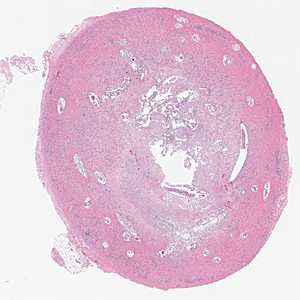
Figure A
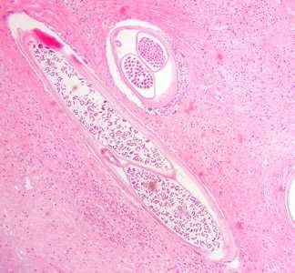
Figure B
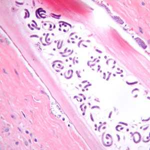
Figure C
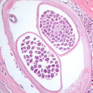
Figure D
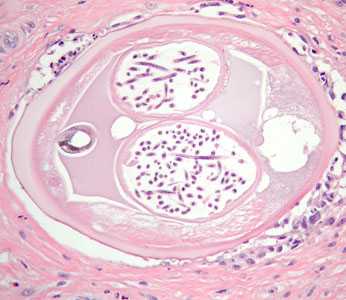
Figure E
Case Answer
This was a case of onchocerciasis caused by Onchocerca volvulus. Diagnostic morphologic features included:
- the presence of adult worms coiled within a subcutaneous nodule (Figure A).
- weak, underdeveloped musculature (MU, Figure E).
- paired uterine tubes with eggs (Figure D) and microfilariae (Figures C and E) in various stages of development. Notice also the small, simple intestine to the left of the uterine tubes in Figures D and E.
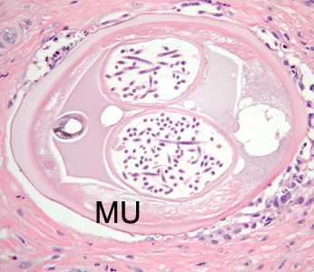
Figure E
More on: onchocerciasis
Images presented in the monthly case studies are from specimens submitted for diagnosis or archiving. On rare occasions, clinical histories given may be partly fictitious.
DPDx is an education resource designed for health professionals and laboratory scientists. For an overview including prevention and control visit www.cdc.gov/parasites/.
- Page last reviewed: August 24, 2016
- Page last updated: August 24, 2016
- Content source:
- Global Health – Division of Parasitic Diseases and Malaria
- Notice: Linking to a non-federal site does not constitute an endorsement by HHS, CDC or any of its employees of the sponsors or the information and products presented on the site.
- Maintained By: