Biology
Causal Agent:
Human baylisascariasis is caused by larvae of Baylisascaris procyonis, an intestinal nematode of raccoons.
Life Cycle:

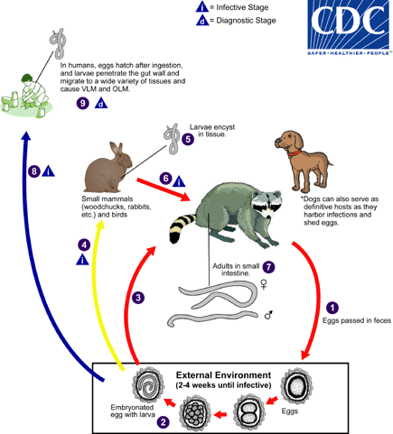
Baylisascaris procyonis completes its life cycle in raccoons, with humans acquiring the infection as accidental hosts (dogs serve as alternate definitive hosts, as they can harbor patent infection and shed eggs). Unembryonated eggs are shed in the environment  , where they take 2-4 weeks to embryonate and become infective
  . Raccoons can be infected by ingesting embryonated eggs from the environment
  . Additionally, over 100 species of birds and mammals (especially rodents) can act as paratenic hosts for this parasite: eggs ingested by these hosts
  hatch and larvae penetrate the gut wall and migrate into various tissues where they encyst
  . The life cycle is completed when raccoons eat these hosts
  . The larvae develop into egg-laying adult worms in the small intestine
  and eggs are eliminated in raccoon feces. Humans become accidentally infected when they ingest infective eggs from the environment; typically this occurs in young children playing in the dirt
  . Migration of the larvae through a wide variety of tissues (liver, heart, lungs, brain, eyes) results in VLM and OLM syndromes, similar to toxocariasis
  . In contrast to Toxocara larvae, Baylisascaris larvae continue to grow during their time in the human host. Tissue damage and the signs and symptoms of baylisascariasis are often severe because of the size of Baylisascaris larvae, their tendency to wander widely, and the fact that they do not readily die. Diagnosis is usually made by serology, or by identifying larvae in biopsy or autopsy specimens.
Life cycle and information courtesy of DPDx.
- Page last reviewed: March 18, 2015
- Page last updated: March 18, 2015
- Content source:


 ShareCompartir