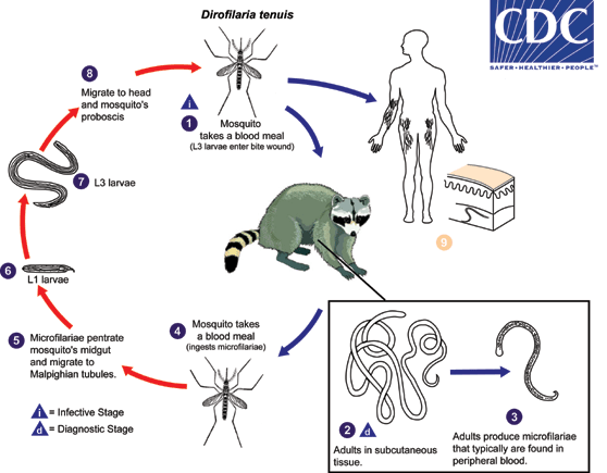
Biology - Life Cycle of D. tenuis
Life Cycle:

During a blood meal, an infected mosquito (Aedes, Anopheles, and possibly others) introduces third-stage filarial larvae of Dirofilaria tenuis onto the skin of the raccoon definitive host (but also occasionally humans), where they penetrate into the bite wound  . In the definitive host, the L3 larvae undergo two more molts into L4 and adults, the latter of which resides in subcutaneous tissues
  . Adult females are usually 80-130 mm long by 260-360 µm wide; males are usually 40-50 mm long by 190-260 µm wide. Adults can live for 5 - 10 years. In subcutaneous tissue, the female worms are capable of producing microfilariae over their lifespan. The microfilariae are found in peripheral blood
  . A mosquito ingests the microfilariae during a blood meal
  . After ingestion, the microfilariae migrate from the mosquito’s midgut through the hemocoel to the Malpighian tubules in the abdomen
  . There the microfilariae develop into first-stage larvae
  and subsequently into third-stage infective larvae
  . The third-stage infective larvae migrate to the mosquito's proboscis
  and can infect another definitive host when it takes a blood meal
  . In humans
  , D. tenuis may wander in the subcutaneous tissues for months, but finally is encapsulated in a granulomatous nodule, but may also be found around the eye or on the conjunctiva. Because of this, the infection in humans was first known as Dirofilaria conjunctivae.
Life cycle image and information courtesy of DPDx.
- Page last reviewed: February 8, 2012
- Page last updated: February 8, 2012
- Content source:


 ShareCompartir