We need you! Join our contributor community and become a WikEM editor through our open and transparent promotion process.
Elbow X-ray
From WikEM
Contents
Four Questions
.png/300px-AnteriorFatPad_(normal).png)
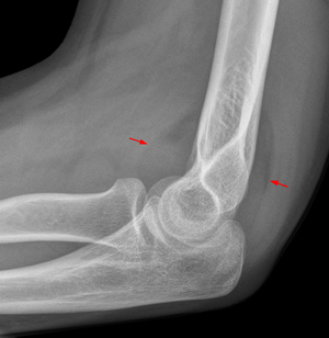
- Are the fat pads normal?
- A visible ant. fat pad is normal but if displaced anteriorly (Sail sign) it is abnormal
- A visible post. fat pad is always abnormal
- What if have fat pad displacement but no fracture or displacement is identified?
- Adults: Treat as radial head fracture
- Peds: Be certain that neither an undisplaced supracondylar fracture nor a displaced internal epicondyle fracture is overlooked!
- Is the radiocapitellar line normal?
- A line drawn along the longitudinal axis of the radial head and neck should pass through the capitellum
- If line does not pass through capitellum then dislocation of radial head is probable
- Whenver there is a fracture of the ulnar shaft must evaluate the radiocapitellar line for possible radial head dislocation (Monteggia fracture dislocation)
- This rule is always valid on a true lateral film
- In pediatric cases the AP view may be misleading
- A line drawn along the longitudinal axis of the radial head and neck should pass through the capitellum
- Is the anterior humeral line normal?
- A line drawn along the ant cortex of the humerus will have at leats 1/3 of the capitellum anterior to it
- If less than 1/3 then strong probability of supracondylar fracture with distal fragment displaced posteriorly
- A line drawn along the ant cortex of the humerus will have at leats 1/3 of the capitellum anterior to it
- Are the ossification centers normal?
- CRITOE (Capitellum, Radial head, Internal epicondyle, Trochlea, Olecranon, Lateral Epicondyle)
- Dislocated elbow may result in avulsion of internal epicondyle
- Because the trochlea ossifies after the internal epicondyle if you see the trochlea you must find the epicondyle!
- Dislocated elbow may result in avulsion of internal epicondyle
- CRITOE (Capitellum, Radial head, Internal epicondyle, Trochlea, Olecranon, Lateral Epicondyle)
See Also
References
- Accident and Emergency Radiology
Video
Authors
Jordan Swartz, Ross Donaldson, Neil Young, Daniel Ostermayer