We need you! Join our contributor community and become a WikEM editor through our open and transparent promotion process.
Myiasis
From WikEM
Contents
Background
- Caused by Diptera species. Dermatobia hominis (botfly) is most common cause in North America
- Cutaneous (includes follicular, wound, and migratory) type is most common
- Can also occur in mouth, urogenital, ophthalmic, nasopharyngeal location
- Typically occurs in tropical and subtropical areas. US cases typically due to travel to endemic region[1]
- Bacterial superinfection is rare[2]
Clinical Features
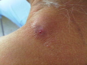
- Erythematous papule with central pore (allows for larval respiration)[2]
- Sensation of movement within lesion
- Serous drainage
Differential Diagnosis
Travel Related Skin Conditions
Papules
- Insect bites
- Scabies
- Seabather's eruption
- Cercarial dermatitis (Swimmer's Itch)
Sub Q Swelling and Nodules
Ulcers
Linear and Migratory Lesions
- Cutaneous Larvae Migrans
- Photodermatitis
Ectoparasites
Evaluation
- Clinical diagnosis
Management[2]
If entire larvae is not removed, severe inflammatory response occurs
- Occlusion of central pore with petroleum jelly or mineral oil interrupts oxygen supply and causes larvae to migrate to surface where it can be grasped with forceps and removed
- Can take up to 24 hours
- Manual removal by squeezing out larvae
- Surgical removal by making incision over larvae and removing with forceps
- Ivermectin - single PO dose or topical application
- Wound myiasis requires surgical debridement
- Ocular, nasopharyngeal, urogenital myiasis should prompt appropriate specialist consultation for management
Disposition
- Cutaneous myiasis generally may be discharged after removal
- Disposition of other forms based on discussion with specialist
See Also
External Links
References
- ↑ https://www.cdc.gov/parasites/myiasis/faqs.html Accessed 02/06/17
- ↑ 2.0 2.1 2.2 McGraw TA. Turiansky GW. Cutaneous myiasis. Journal of the American Academy of Dermatology. 58(6):907-26; quiz 927-9, 2008 Jun.
Authors
Michael Holtz, Ross Donaldson, Neil Young, Daniel Ostermayer

