We need you! Join our contributor community and become a WikEM editor through our open and transparent promotion process.
Necrotizing fasciitis
From WikEM
Contents
Background
- A rapidly progressive infection primarily involving the fascia and subcutaneous tissue
- Formerly a rare diagnosis, frequency has risen due to an increase in immunocompromised patients with significant risk factors[1]
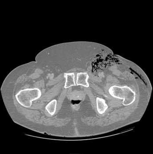
- Gas-formation is NOT a requirement for diagnosis, and radiographical lack of the classically taught gas formation should NEVER rule out necrotizing infection[2]
- Most severe form of soft tissue infection and potentially limb and life threatening
- Early recognition and aggressive debridement are major prognostic determinants and delay increases mortality[3]
Categories
- Type I, polymicrobial
- Type II, group A streptococcal
- Type III, gas gangrene or clostridial myonecrosis
Risk Factors
- DM
- Drug use
- Obesity
- Immunosuppression
- Recent surgery
- Traumatic wounds
Clinical Features
- Skin exam
- Erythema (without sharp margins)
- Exquisitely tender (pain out of proportion to exam)
- Caveat - some patients present with "la belle indifference"
- May be a result of ischemic, insensate tissue[4]
- Skip lesions
- Hemorrhagic bullae (violaceous bullae)
- May be preceded by skin anesthesia (destruction of superficial nerves)
- Crepitus (in type I infections)
- Swelling/edema may produce compartment syndrome
- Constitutional
- Fever
- Tachycardia
- Systemic toxicity
Differential Diagnosis
Skin and Soft Tissue Infection
- Cellulitis
- Erysipelas
- Lymphangitis
- Abscess
- Necrotizing soft tissue infections
- Necrotizing fasciitis
- Necrotizing myositis
- Necrotizing cellulitis
- Mycobacterium marinum
Look-A-Likes
Necrotizing rashes
- Necrotizing soft tissue infections
- Necrotizing fasciitis
- Necrotizing myositis
- Necrotizing cellulitis
- Purpura fulminans
- Drug rash
- Levamisole toxicity
Evaluation
Work-Up
- CBC
- Chem
- PT/PTT/INR
- CK
- Lactate
Evaluation
- Surgical exploration is the ONLY way to definitively establish the diagnosis of necrotizing infection
- Imaging
- Should not delay surgical exploration
- CT is study of choice - soft tissue gas, edema and fluid collections, fascial thickening with fat stranding
- US may show thickened fascial planes, fluid between fascial planes, irregularity of the fascia, subcutaneous emphysema. The study may be limited by soft tissue gas
- MRI - T2 subcutaneous, intramuscular, and fascial edema
Laboratory Risk Indicator for Necrotizing Fasciitis (LRINEC) Score[5]
Has not been prospectively validated, index of suspicion is key and 10% of the patients with a score < 6 had Necrotizing Fasciitis. A score > 6 has PPV of 92% and NPV of 96% for necrotizing fasciitis.
- CRP (mg/L) ≥150: 4 points
- WBC count (×103/mm3)
- <15: 0 points
- 15–25: 1 point
- >25: 2 points
- Hemoglobin (g/dL)
- >13.5: 0 points
- 11–13.5: 1 point
- <11: 2 points
- Sodium (mmol/L) <135: 2 points
- Creatinine (umol/L) >141: 2 points
- Glucose >180 mg/dL (10 mmol/L): 1 point
Grouping by Scores
- Low Risk: score 5 (10% of pts with score < 6 still had nec fasc)
- Moderate Risk: score 6– 7
- High Risk: score >8
HUCLA NF vs Non-NF Criteria:[6]
- Retrospective study discovered:
- WBC count >15.4(x103/mm3) OR Na <135(mmol/L)
- Associated with NF and combo of both increased likelihood of NF
- PPV 26%/NPV 99%
- Useful tool to rule out NF, not a good tool for confirming presence of NF
- Helps distinguish NF from non-NF infection, when classic 'hard' signs of NF are absent however clinical judgment should still be used in patient with high suspicion of the disease
Management
- Surgical exploration and debridement is both the definitive diagnostic modality and the definitive treatment
- Indicated in setting of severe pain, toxicity, fever, elevated CK (with or without radiographic evidence)
- Antibiotics
- Must cover gram positives, gram negatives, and anaerobes (especially GAS and clostridium)
- Piperacillin-Tazobactam 3.375-4.5g q6hr AND clindamycin 600-900mg q8hr AND vancomycin 1gm IV q12hr (consider weight base dosing of 20 mg/kg)
- In diabetics, maintain strict glycemic control (with IVFs and IV insulin if necessary)
Disposition
- Admit to ICU
See Also
- Necrotizing soft tissue infections
- Laboratory Risk Indicator for Necrotizing Fasciitis (LRINEC) Score
- EBQ:LRINEC Score
Video
References
- ↑ Hakkarainen TW et al. Necrotizing soft tissue infections: review and current concepts in treatment, systems of care, and outcomes. Curr Probl Surg. 2014 Aug. 51 (8):344-72.
- ↑ Misiakos EP et al. Current concepts in the management of necrotizing fasciitis. Front Surg. 2014. 1:36.
- ↑ Wong CH, Khin LW, Heng KS, Tan KC, Low CO (2004). "The LRINEC Laboratory Rother soft tissue infections". Critical Care Medicine 32 (7): 1535–1541. doi:10.1097/01.CCM.0000129486.35458.7D. PMID 15241098
- ↑ TheHealthScience. Emergent Management of Necrotizing Soft Tissue Skin Infections. Nov 22, 2013. https://thehealthscience.com/topics/emergent-management-necrotizing-soft-tissue-skin-infections.
- ↑ Wong C. "The LRINEC (Laboratory Risk Indicator for Necrotizing Fasciitis) score: a tool for distinguishing necrotizing fasciitis from other soft tissue infections". Crit Care Med. 2004. 32(7):1535-41.
- ↑ Wall DB et al. A simple model to help distinguish necrotizing fasciitis from nonnecrotizing soft tissue infection. J Am Coll Surg. 2000 Sep;191(3):227-31.


