We need you! Join our contributor community and become a WikEM editor through our open and transparent promotion process.
File list
This special page shows all uploaded files.
First page |
Previous page |
Next page |
Last page |
| Date | Name | Thumbnail | Size | User | Description | Versions |
|---|---|---|---|---|---|---|
| 07:31, 11 May 2017 | Teolipic.jpg (file) |  |
168 KB | Dteoli | Dac Teoli | 2 |
| 00:51, 10 May 2017 | Purple glove syndrome.png (file) |  |
347 KB | Lisayee25 | Senthilkumaran S, Balamurgan N, Suresh P, Thirumalaikolundusubramanian P. Purple glove syndrome: A looming threat. J Neurosci Rural Pract. 2010;1(2):121-2. | 1 |
| 04:01, 6 May 2017 | PMC3522363 iranjradiol-08-205-g002.png (file) | 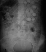 |
142 KB | Lisayee25 | Shahnazi M, Sanei taheri M, Pourghorban R. Body packing and its radiologic manifestations: a review article. Iran J Radiol. 2011;8(4):205-10. | 1 |
| 22:25, 28 April 2017 | Posthipdislocation.jpg (file) | 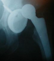 |
19 KB | Rossdonaldson1 | By Cindy Funk - http://www.flickr.com/photos/cindyfunk/91201732/, CC BY 2.0, https://commons.wikimedia.org/w/index.php?curid=15450352 | 1 |
| 22:23, 28 April 2017 | HipdisX.png (file) | 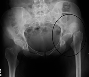 |
723 KB | Rossdonaldson1 | By James Heilman, MD - Own work, CC BY-SA 3.0, https://commons.wikimedia.org/w/index.php?curid=16263165 | 1 |
| 21:42, 28 April 2017 | Patellaluxation ap 001.png (file) | 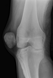 |
490 KB | Rossdonaldson1 | By Hellerhoff - Own work, CC BY-SA 3.0, https://commons.wikimedia.org/w/index.php?curid=11989009 | 1 |
| 21:32, 28 April 2017 | AnterDisMark.png (file) | 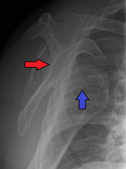 |
715 KB | Rossdonaldson1 | By James Heilman, MD - Own work, CC BY-SA 4.0, https://commons.wikimedia.org/w/index.php?curid=49111467 | 1 |
| 21:31, 28 April 2017 | AnterDisAPMark.png (file) | 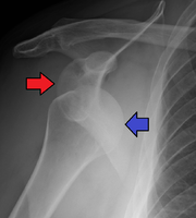 |
671 KB | Rossdonaldson1 | By James Heilman, MD - Own work, CC BY-SA 4.0, https://commons.wikimedia.org/w/index.php?curid=49111463 | 1 |
| 21:22, 28 April 2017 | Luxation epaule.png (file) | 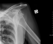 |
97 KB | Rossdonaldson1 | By MB - Collection personnelle, CC BY-SA 2.5, https://commons.wikimedia.org/w/index.php?curid=1254054 | 1 |
| 21:19, 28 April 2017 | Inferiourdislocation.jpg (file) | 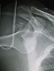 |
752 KB | Rossdonaldson1 | By James Heilman, MD - Own work, CC BY-SA 3.0, https://commons.wikimedia.org/w/index.php?curid=9500637 | 1 |
| 21:13, 28 April 2017 | Splint1.jpg (file) | 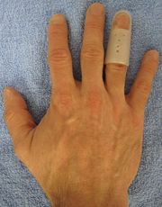 |
122 KB | Rossdonaldson1 | By James Heilman, MD - Own work, CC BY-SA 3.0, https://commons.wikimedia.org/w/index.php?curid=12789324 | 1 |
| 20:56, 28 April 2017 | Mallet finger.png (file) | 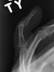 |
46 KB | Rossdonaldson1 | By James Heilman, MD - Own work, CC BY-SA 3.0, https://commons.wikimedia.org/w/index.php?curid=12179191 | 1 |
| 20:55, 28 April 2017 | MalletFinger.png (file) | 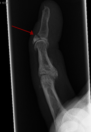 |
263 KB | Rossdonaldson1 | By James Heilman, MD - Own work, CC BY-SA 3.0, https://commons.wikimedia.org/w/index.php?curid=12496361 | 1 |
| 20:52, 28 April 2017 | Mallet finger.jpg (file) | 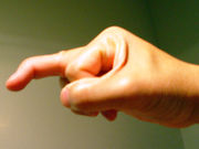 |
227 KB | Rossdonaldson1 | By Howcheng - Own work, CC BY-SA 3.0, https://commons.wikimedia.org/w/index.php?curid=4422969 | 1 |
| 18:10, 28 April 2017 | Tape15.png (file) | 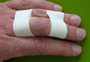 |
61 KB | Rossdonaldson1 | By Redlinux - Own work, CC BY 3.0, https://commons.wikimedia.org/w/index.php?curid=11419281 | 1 |
| 18:07, 28 April 2017 | Lightbulb sign - posterior shoulder dislocation - Roe vor und nach Reposition 001.jpg (file) | 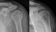 |
96 KB | Rossdonaldson1 | By Hellerhoff - Own work, CC BY-SA 3.0, https://commons.wikimedia.org/w/index.php?curid=39582858 | 1 |
| 18:05, 28 April 2017 | Dislocated Finger.jpg (file) | 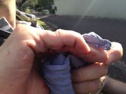 |
126 KB | Rossdonaldson1 | By Mdumont01 - Own work, CC BY-SA 3.0, https://commons.wikimedia.org/w/index.php?curid=30083592 | 1 |
| 17:57, 28 April 2017 | MCCdislocation.png (file) | 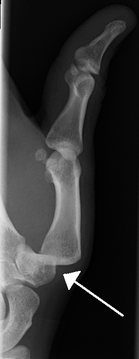 |
145 KB | Rossdonaldson1 | By James Heilman, MD - Own work, CC BY-SA 3.0, https://commons.wikimedia.org/w/index.php?curid=14634845 | 1 |
| 17:49, 28 April 2017 | Ankledislocation.jpg (file) | 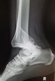 |
1,008 KB | Rossdonaldson1 | By James Heilman, MD - Own work, CC BY-SA 3.0, https://commons.wikimedia.org/w/index.php?curid=11110496 | 1 |
| 17:45, 28 April 2017 | Dislocated Finger XRay.png (file) | 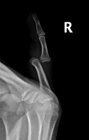 |
118 KB | Rossdonaldson1 | By Mdumont01 - Own work, CC BY-SA 3.0, https://commons.wikimedia.org/w/index.php?curid=30083608 | 1 |
| 11:46, 27 April 2017 | PMC2892654 CRM2010-213818.004.png (file) |  |
158 KB | Rossdonaldson1 | OPENi Acute tension pneumothorax following cardiac herniation after pneumonectomy. Steinmann D, Rohr E, Kirschbaum A - Case Rep Med (2010) Bottom Line: A tension pneumothorax is one of the main causes of cardiac arrest in the initial postoperative period after thoracic surgery.Tension pneumothorax and cardiac herniation must be taken into account in hemodynamically unstable patients after pneumonectomy.We report an unusual case of successful treatment of acute tension pneumothorax following cardiac herniation and intrathoracic bleeding after pneumonectomy. View Article: PubMed Central - PubMed Affiliation: Department of Anaesthesia and Critical Care Medicine, University Hospital Freiburg, Hugstetter Strasse 55, 79106 Freiburg, Germany. | 1 |
| 09:14, 27 April 2017 | Radiograph of Barton's fracture.jpg (file) |  |
57 KB | Rossdonaldson1 | 1 | |
| 03:50, 22 April 2017 | Red Eye - Advanced.png (file) | 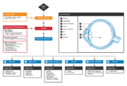 |
578 KB | Tomfadial | {{WikEM algorithm project}} Link: https://www.lucidchart.com/documents/edit/0cbb6a6e-c89c-4e43-9f77-27f68c6d905e | 1 |
| 12:39, 21 April 2017 | Lund-Browder chart-burn injury area.PNG (file) |  |
32 KB | Neil.m.young | Lund-Browder Chart Wikicommons | 1 |
| 21:18, 14 April 2017 | Erythema toxcium.png (file) | 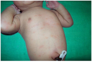 |
364 KB | Stcmd | https://openi.nlm.nih.gov/detailedresult.php?img=PMC3918370_ISRN.DERMATOLOGY2014-360590.010&query=erythema+toxicum&it=xg&req=4&npos=1 | 1 |
| 14:23, 10 April 2017 | TFan.jpg (file) |  |
837 KB | Ted Fan | profile | 1 |
| 01:59, 5 April 2017 | Opioid abuse graph.png (file) | 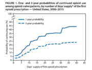 |
44 KB | Kxl328 | 1 | |
| 21:10, 3 April 2017 | Legs over head PSVT.jpeg (file) |  |
32 KB | Kxl328 | 1 | |
| 15:50, 3 April 2017 | Gray810.png (file) | 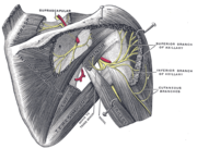 |
52 KB | Rossdonaldson1 | By Henry Vandyke Carter - Henry Gray (1918) Anatomy of the Human Body (See "Book" section below)Bartleby.com: Gray's Anatomy, Plate 810, Public Domain, https://commons.wikimedia.org/w/index.php?curid=541666 | 1 |
| 05:59, 22 March 2017 | Riverside.jpg (file) |  |
13 KB | Dteoli | ../images/New-Tower-Image-1-300px.jpg | 1 |
| 17:42, 18 March 2017 | Spectrum.png (file) | 23 KB | Tomfadial | Blunt Cardiac Injury Spectrum | 1 | |
| 08:01, 15 March 2017 | Pediatric vs adult LP.jpg (file) | 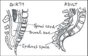 |
76 KB | Kxl328 | 1 | |
| 07:58, 15 March 2017 | Pediatric spinal cord.JPG (file) |  |
33 KB | Kxl328 | 1 | |
| 00:06, 10 March 2017 | Hemocue.png (file) |  |
64 KB | Tomfadial | 1 | |
| 03:03, 28 February 2017 | AVD without CHB.png (file) |  |
193 KB | Kxl328 | 1 | |
| 00:37, 27 February 2017 | Ddx for jaundice by labs.gif (file) |  |
7 KB | Kxl328 | 1 | |
| 00:02, 27 February 2017 | Ino v2.png (file) | 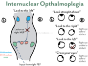 |
191 KB | Kxl328 | 1 | |
| 07:21, 20 February 2017 | Takayasu MRA.png (file) | 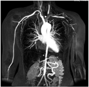 |
226 KB | Devin Smith | MRA of a patient with significant vascular narrowing from Takayasu Arteritis Takayasu arteritis: criteria for surgical intervention should not be ignored. Perera AH, Mason JC, Wolfe JH - Int J Vasc Med (2013) Open Access | 1 |
| 23:05, 19 February 2017 | Nerves of the left upper extremity.gif (file) |  |
112 KB | Devin Smith | From Wikipedia | 1 |
| 22:21, 19 February 2017 | Ulnar Elbow.jpg (file) | 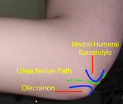 |
699 KB | Devin Smith | Elbow showing ulnar nerve pathway | 1 |
| 12:29, 15 February 2017 | Liquid Pool Chlorine.jpg (file) |  |
57 KB | Rossdonaldson1 | By Maksym Kozlenko - Own work, CC BY-SA 4.0, https://commons.wikimedia.org/w/index.php?curid=44997263 | 1 |
| 11:12, 15 February 2017 | Piles Grade 4.svg (file) |  |
219 KB | Rossdonaldson1 | By Armin Kübelbeck, CC BY 3.0, https://commons.wikimedia.org/w/index.php?curid=18552387 | 1 |
| 11:11, 15 February 2017 | Piles Grade 3.svg (file) |  |
208 KB | Rossdonaldson1 | By Armin Kübelbeck, CC BY 3.0, https://commons.wikimedia.org/w/index.php?curid=18552385 | 1 |
| 11:11, 15 February 2017 | Piles 4th deg 01.jpg (file) | 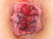 |
63 KB | Rossdonaldson1 | By Dr. K.-H. Günther, Klinikum Main Spessart, Lohr am Main, CC BY 3.0, https://commons.wikimedia.org/w/index.php?curid=20649949 | 1 |
| 11:10, 15 February 2017 | Hemrrhoids 05.jpg (file) | 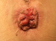 |
292 KB | Rossdonaldson1 | By Prof. Dr. A. Herold, End- und Dickdarm-Zentrum Mannheim, CC BY 3.0, https://commons.wikimedia.org/w/index.php?curid=18924452 | 1 |
| 11:09, 15 February 2017 | Hemrrhoids 04.jpg (file) | 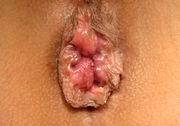 |
891 KB | Rossdonaldson1 | By Prof. Dr. A. Herold, End- und Dickdarm-Zentrum Mannheim, CC BY 3.0, https://commons.wikimedia.org/w/index.php?curid=18924444 | 1 |
| 11:09, 15 February 2017 | Piles Grade 2.svg (file) |  |
192 KB | Rossdonaldson1 | By Armin Kübelbeck, CC BY 3.0, https://commons.wikimedia.org/w/index.php?curid=18552373 | 1 |
| 11:08, 15 February 2017 | Haemorrhoiden 1Grad endo 01.jpg (file) | 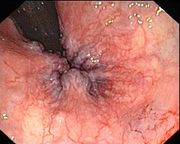 |
223 KB | Rossdonaldson1 | By Dr. Joachim Guntau - www.Endoskopiebilder.de, CC BY-SA 3.0, https://commons.wikimedia.org/w/index.php?curid=18660115 | 1 |
| 11:07, 15 February 2017 | Piles Grade 1.svg (file) |  |
174 KB | Rossdonaldson1 | By Armin Kübelbeck, CC BY 3.0, https://commons.wikimedia.org/w/index.php?curid=18552361 | 1 |
| 11:04, 15 February 2017 | Perianal thrombosis 01.jpg (file) | 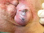 |
62 KB | Rossdonaldson1 | By Dr. K.-H. Günther, Klinikum Main Spessart, Lohr am Main, CC BY 3.0, https://commons.wikimedia.org/w/index.php?curid=20649959 | 1 |
First page |
Previous page |
Next page |
Last page |
