We need you! Join our contributor community and become a WikEM editor through our open and transparent promotion process.
Testicular ultrasound
From WikEM
Contents
Background
- Testicular torsion is a medical emergency where acute treatment is needed
- Testicular salvage rate by time[1]
- Near 100% within 6 hours
- 70% within 6-12 hours
- 20% within 12-24 hours
Anatomy
- Testicle - ~2 to 3 cm in width and 3 to 5 cm in length
- Epididymis - along the posterolateral aspect of each testis
- Vas Deferens
- Spermatic cord
- Median Raphe
Indications
- Testicular pain or tenderness
- Testicular swelling
Technique
- Select probe
- Linear probe
- Location
- Obtain anterior views over the scrotum
- Landmarks
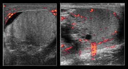
- Heterogeneous testicles can be seen under the soft tissues of the scrotum
- Obtain sagittal and transverse images
- Initial view should be “buddy view” identifying both testicles in a tranverse plane with the middle of the probe over the scrotal raphe
- Each testicle should be examined in the transverse and sagittal plane
- Each testicle should be examined under power Doppler assessing blood flow
- Optimize image quality
- Minimize depth to include only the body of the testicle
- When assessing blood flow, increase power Doppler to a level where vasculature can be easily identified
Findings
Normal Testicle
- Equal heterogeneity
- Equal blood flow
- Minimal fluid around the testicle
Testicular Torsion
- Enlarged testicle
- Increased heterogeneity
- Decreased flow on power Doppler
- Increased flow may be seen in cases of torsion with subsequent detorsion
Epididymitis
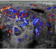
- Enlarge epididymis
- Increased epididymal blood flow
- Normal testicular appearance
Hydrocele
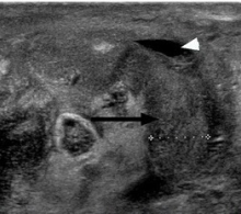
- Anechoic fluid adjacent to the testicle
Orchitis
- Enlarged testicle
- Increased flow on power Doppler
- Difficult to differentiate from recently detorsed testicle
Images
Normal
Abnormal
Testicular Torsion
Epididymitis
Hydrocele
Pearls and Pitfalls
- Adjust color flow on normal testicle first as a baseline to compare to painful or swollen testicle
- Hyperemic testicle does not rule out testicular torsion as this could represent intermittent torsion or detorsion
Documentation
Normal Exam
A bedside ultrasound was conducted to assess for signs testicular torsion with clinical indications of right/left testicular pain. The right and left testicles were identified and viewed in the transverse and sagittal plane and assessed with power Doppler. Heterogenicity was similar and blood flow was similar. There was not indications of testicular torsion.
Abnormal Exam
A bedside ultrasound was conducted to assess for signs testicular torsion with clinical indications of right/left testicular pain. The right and left testicles were identified and viewed in the transverse and sagittal plane and assessed with power Doppler. The right/left testicle has increased heterogenicity and diminished blood flow. Findings indicate testicular torsion.
Clips
Normal
Abnormal
Testicular Torsion
External Links
See Also
References
- ↑ Patriquin HB, Yazbeck S, Trinh B, et al. Testicular torsion in infants and children: diagnosis with Doppler sonography. Radiology. 1993;188:781-785.


