We need you! Join our contributor community and become a WikEM editor through our open and transparent promotion process.
Ultrasound: Joint
From WikEM
Contents
Background
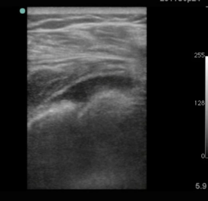
- U/S can demonstrate joint effusions and aid in diagnostic procedures such as joint aspiration and hematoma blocks
- Common joints that are amenable to ultrasound includes glenohumoral, elbow, wrist, hip, knee, and ankle joint
- Shoulder dislocations can be found with SN and SP nearing 100% in both diagnosis and assessing reduction[1]
Joint Effusion
Images
Normal
Abnormal
Instructions
- Select linear probe (high frequency probe) for smaller joints such as elbow, wrist, ankle, or knee
- Select curvelinear probe (low frequency probe) for larger joints such as shoulder or hip joint
- Scan joint longitudinally to identify the joint space and adjacent bones
- Rotate 90° over area of concern
Findings
- Positive
- Substantial quantity of anechoic fluid (in comparison to contralateral side)
- Negative
- Trace or no effusion
Pearls and Pitfalls
- Compare contralateral joint
Shoulder Dislocation
Images
Normal
Abnormal
Instructions
Posterior Approach
- Select curvilinear probe (low frequency probe)
- Place probe to the posterior chest parallel above the scapular spine
- Identify the glenoid and humeral head
Anterior Approach
- Select curvilinear probe (low frequency probe)
- Place probe to the anterior chest parallel to the glenohumeral joint
- Identify the glenoid and humeral head
Findings
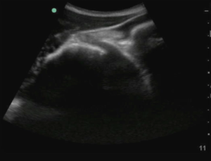
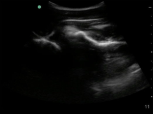
- In anterior and posterior shoulder dislocations, the humeral head is displaced anterior or posterior to the glenoid fossa, respectively
- Hematoma may be present and appear hyperechoic next to the glenoid fossa
Pearls and Pitfalls
- Always compare findings with the unaffected joint
See Also
External Links
References
- ↑ Abbasi, S, et al. Diagnostic Accuracy of Ultrasonographic Examination in the Management of Shoulder Dislocation in the Emergency Department. Annals of Emergency Medicine. 2013; 62(2):170–175.


