Ocular ultrasound
Contents
Background
- U/S has been well established in the ophtho literature but new advent to ED practice
Indications
- Acute/Subacute vision changes or loss
- Symptoms of increased ICP
- Suspect FB or globe rupture
- Trauma (assess for reactive pupils)
Technique
- Patient preparation
- Apply large transparent film dressing (such as Tegaderm) over the closed eye taking care not to create air bubbles between the film and skin surface
- Deposit a copious amount of ultrasound gel to the film
- Select probe
- Linear probe
- Location
- Probe should be applied to the gel without creating significant pressure on the globe itself (especially in situations of suspected globe rupture)
- Landmarks
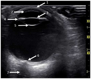
- The round structure of the globe should be easily identified with the lens in the anterior section
- Gain should be increased significantly to the point where echos are generating within the vitreous humor
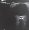
- If needed, measurements of the optic nerve can be taken as still image
Findings
Elevated ICP
- Measure optic nerve 3mm posterior to the globe/papilla, from inner wall to inner wall
- Normal is <5mm[1]
- U/S has a sensitivity of 95.6% and specificity of 92.3% for increased ICP[2]
Globe Rupture
- Only perform if you can ensure that you do not put pressure on the globe
- Findings
- Decrease in size of globe
- Anterior chamber collapse
- Vitreous hemorrhage
- Buckling of the sclera
Intraocular Foreign Body
- Bright, echogenic acoustic profile with associated shadowing or reverberation
Retinal Detachment
- Echogenic undulating membrane in the posterior globe, protruding into the vitreous
- Evaluate with patient moving eye left/right
- SN 97-100% and SP 83-100%[3]
Vitreous Hemorrhage
- Vitreous filled with multiple large echoes
- Increasing the gain is helpful for detecting acute hemorrhages
Images
Normal
- Eyelid
- Cornea
- Lens
- Ciliary bodies
- Lens
- Vitreous chamber
- Optic nerve
Abnormal
Pearls and Pitfalls
- Use copious amount of gel to prevent the need for applying excessive pressure
- A high gain is generally required in order to see vitreous and retinal pathology
- Many findings are more apparent with active eye movement
Documentation
Normal Exam
A bedside ultrasound was conducted to assess for signs of retinal detachment or vitreous hemorrhage with clinical indications of left/right sided vision changes. The anterior chamber, lens, and posterior chamber of the left/right eye was identified during active movement. There was no evidence of retinal detachment or vitreous hemorrhage found. There was no sonographic evidence of retinal detachment or vitreous hemorrhage.
Abnormal Exam
A bedside ultrasound was conducted to assess for signs of retinal detachment or vitreous hemorrhage with clinical indications of left/right sided vision changes. The anterior chamber, lens, and posterior chamber of the left/right eye was identified during active movement. There was a hyperechoic flap located in the anterior chamber/swirling debris in the posterior chamber. There was sonographic evidence of retinal detachment/vitreous hemorrhage.
Clips
Normal
Abnormal
External Links
See Also
Video
References
- ↑ Blaivas M, Theodoro D, Sierzenski PR. Elevated intracranial pressure detected by bedside emergency ultrasonography of the optic nerve sheath. Acad Emerg Med. 2003 Apr;10(4):376-81.
- ↑ Ohle R, McIsaac SM, Woo MY, et al. Sonography of the Optic Nerve Sheath Diameter for Detection of Raised Intracranial Pressure Compared to Computed Tomography: A Systematic Review and Meta-analysis. J Ultrasound Med. 2015; 34(7):1285-1294.
- ↑ Vrablik, ME, et al. The Diagnostic Accuracy of Bedside Ocular Ultrasonography for the Diagnosis of Retinal Detachment: A Systematic Review and Meta-analysis. Annals of Emergency Medicine. 2015; 65(2):199–203.e1.


