We need you! Join our contributor community and become a WikEM editor through our open and transparent promotion process.
Ultrasound: Tendons
From WikEM
Contents
Background
- U/S can be used to assess continuity of tendons and ligaments
- They have a property called anisotropy which means they have 2 different appearances if assess longitudinally or transversely
Images
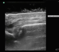
Normal
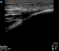
Abnormal
Instructions
- Use linear probe (high frequency probe)
- Place probe in longitudinal plane over suspect tendon; high yield locations inlcude:
- Biceps can be proximal or distal
- Patella tendons 2cm from insertion on patella
- Achilles 2-6cm above calcaneus
- Fan and slide side to side to optimize your view
- Slide distal to proximal to find defect
- Turn probe 90° to assess for tendon body defects
Findings
- Positive Findings
- Discontinuity in longitudinal view of ligament
- Collection of fluid in longitudinal or transverse view suggests injury
- Negative Findings
- Longitudinal views show continuous densely striped parallel lines
- Transverse views show oval structure with punctate interior
Pearls and Pitfalls
- Look at other limb for "normal" anatomy
- Have patient range limb and view in real time
- Know your limitations if case is not clear cut
- Do not mistake anisotropy for tendon rupture


