
Fleas
[Ctenocephalides canis] [Ctenocephalides felis] [Pulex irritans] [Xenopsylla cheopis]
Causal Agents
Many species of fleas can feed on humans. The human flea, Pulex irritans, is less-commonly seen these in industrialized areas. This species is not an effective vector of disease but can serve as an intermediate host for the cestodes Dipylidium caninum and Hymenolepis nana. The cat and dog fleas (Ctenocephalides canis and C. felis) may also feed on humans. In addition to being intermediate hosts for cestodes, C. felis can serve as a vector of Bartonella henselae (cat-scratch disease) and Rickettsia felis (feline rickettsiae). The Oriental rat flea (Xenopsylla cheopis) is the primary vector for Yersinia pestis (plague). Humans with close contact with birds may also be fed upon by the sticktight flea (Echidnophaga gallinacea). The chigo flea (Tunga penetrans) is discussed separately here.
General Flea Life Cycle
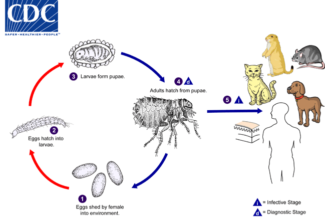
Fleas, like other holometabolous insects, have a four-part life cycle consisting of eggs, larvae, pupae, and adults. Eggs are shed by the female in the enviroment  . Eggs hatch into larvae
  in about 3-4 days and feed on organic debris in the environment. The number of larval instars varies among the species. Larvae eventually form pupae
  , which are in cocoons that are often covered with debris from the environment (sand, pebbles, etc). The larval and pupal stages take about 3-4 weeks to complete. Afterwards, adults hatch from pupae
  and seek out a warm-blooded host for blood meals. The primary hosts for Ctenocephalides felis and C. canis are cats and dogs, respectively, although other mammals, including humans, may be fed upon. The primary hosts for Xenopsylla cheopis are rodents, especially rats. In North America, plague (Yersinia pestis) is cycled between X. cheopis and prairie dogs. Humans are the primary host for Pulex irritans.
The chigoe flea (Tunga penetrans) has different life cycle and is discussed in more detail here.
Geographic Distribution
Worldwide.
Ctenocephalides canis and C. felis.
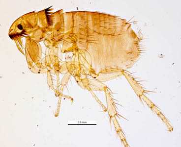
Figure A: The cat flea, C. felis. Image courtesy of Parasite and Diseases Image Library, Australia.
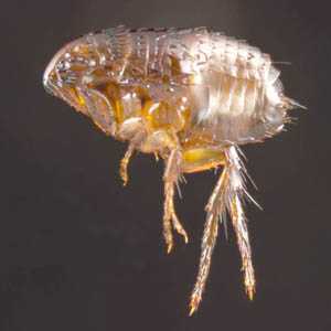
Figure B: The cat flea, C. felis. Image courtesy of Parasite and Diseases Image Library, Australia.
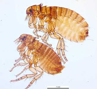
Figure C: The dog flea, C. canis. Image courtesy of Parasite and Diseases Image Library, Australia.
Pulex irritans.
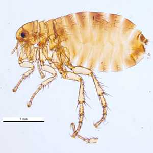
Figure A: The human flea, P. irritans. Image courtesy of Parasite and Diseases Image Library, Australia.
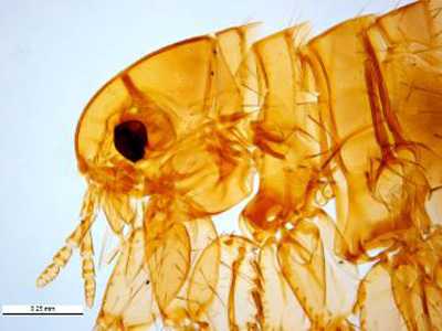
Figure B: The human flea, P. irritans. This image shows a close-up of the head region; note a lack of genal and pronotal combs. Image courtesy of Parasite and Diseases Image Library, Australia.
Xenopsylla cheopis.
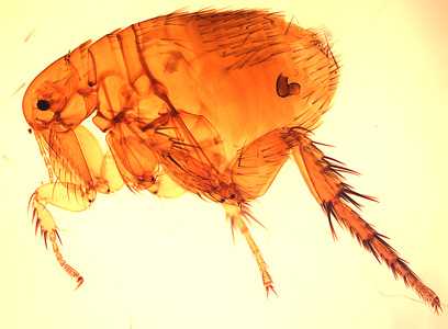
Figure A: The Oriental rat flea, Xenopsylla cheopis.
DPDx is an education resource designed for health professionals and laboratory scientists. For an overview including prevention and control visit www.cdc.gov/parasites/.
- Page last reviewed: May 3, 2016
- Page last updated: May 3, 2016
- Content source:
- Global Health – Division of Parasitic Diseases and Malaria
- Notice: Linking to a non-federal site does not constitute an endorsement by HHS, CDC or any of its employees of the sponsors or the information and products presented on the site.
- Maintained By:


 ShareCompartir