We need you! Join our contributor community and become a WikEM editor through our open and transparent promotion process.
Elbow dislocation
From WikEM
Contents
Background
- Usually due to FOOSH
- 90% are posterolateral
- Median and ulnar nerves may be injured
- "Terrible Triad" injury describes unstable joint consisting of:
- Elbow dislocation
- Radial head fracture
- Coronoid fracture
Clinical Features
- Swelling may be severe
- Displaced equilateral triangle of olecranon and epicondyles (undisturbed in supracondylar fracture)
Posterior dislocation
- Elbow held in 45 degree of flexion
- Olecranon is prominent posteriorly
Anterior dislocation
- Elbow held in extension
Differential Diagnosis
Elbow Diagnoses
Radiograph-Positive
- Radial head fracture
- Capitellum fracture
- Olecranon fracture
- Elbow dislocation
Radiograph-Negative
- Lateral epicondylitis
- Medial epicondylitis
- Olecranon bursitis (nonseptic)
- Septic bursitis
- Biceps tendon rupture/dislocation
Pediatric
- Nursemaid's elbow
- Supracondylar fracture
- Lateral epicondyle fracture
- Medial epicondyle fracture
- Olecranon fracture
- Radial head fracture
- Salter-Harris fractures
Evaluation
- Imaging
- Look for associated fractures (especially of coronoid and radial head)
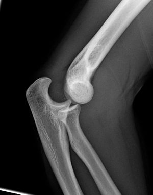
- Lateral: both ulna and radius are displaced posteriorly
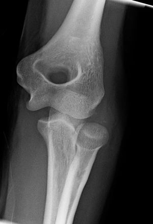
- AP: lateral or medial displacement with ulna/radius in their normal relationship
- Red flags
- Compartment syndrome
- Neurovascular injury
- Open dislocations
Management
- Likely require Procedural sedation
- Reduction techniques: [1]
- Longitudinal traction on wrist/forearm with downward pressure on forearm
- Patient lies prone
- Assistant pulls counter traction on humerus
- Provider pulls longitudinally with elbow in extension, then flexes elbow
- Stimson
- Patient prone with elbow flexed at 90 degrees at edge of bed. Hang weight from hand, and if needed provider can push olecranon into place
- Immobilize in long arm posterior mold with elbow in slightly less than 90deg flexion
- If unstable, splint with forearm in pronation
- Document post reduction neurovascular status and post reduction films
Disposition
- Obtain emergent consult for irreducible dislocations, nerve or vascular compromise, associated fracture, open dislocation
- Simple dislocation requires ortho follow up within 1 week
See Also
References
Cite error: <ref> tags exist, but no <references/> tag was found


