We need you! Join our contributor community and become a WikEM editor through our open and transparent promotion process.
Meniscus and ligament knee injuries
From WikEM
Contents
Background
- Anterior Cruciate Ligament
- Limits anterior translation of tibia
- 75% of all hemarthroses are caused by disruption of ACL
- Posterior Cruciate Ligament
- Limits posterior translation of tibia
- Isolated injuries are rare
- Medial Collateral Ligament
- Provide restraint against valgus (outward) stress
- Lateral Collateral Ligament
- Provide restraint against varus (inward) stress
Clinical Features
ACL
- Hearing/feeling a "pop" during injury with ensuing knee instability is pathognomonic
- Anterior Drawer Sign
- Pt supine, knee flexed 90', attempt to displace tibia from femur in a forward direction
- Displacement of >6mm compared w/ opposite knee indicates injury
- Lachman Test (most sensitive)
- Pt supine, knee flexed 30', femur held w/ one hand, prox tibia pulled up w/ other hand
- Displacement >5mm or soft end-point indicates injury
- Pivot Shift Test
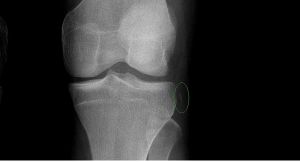
- Segond Fracture
- Pathognomonic for ACL tear but rare
PCL
- Posterior Drawer Sign
- Patient supine, knee flexed 90', attempt to displace tibia from femur in backward direction
Meniscus
- Symptoms
- "Locking" of joint or sensation of popping, clicking, or snapping
- Signs
- Effusions that occur after activity
- Joint-line tenderness
- Tests
- McMurray, grind test only 50% Sn
Differential Diagnosis
Knee diagnoses
Acute Injury
- Knee fractures
- Patella fracture
- Tibial plateau fracture
- Knee dislocation
- Patella dislocation
- Segond fracture
- Meniscus and ligament knee injuries
- Patellar Tendinitis (Jumper's knee)
- Patellar tendon rupture
- Quadriceps tendon rupture
Nontraumatic/Subacute
- Septic Joint
- Gout
- Popliteal cyst (Baker's)
- Prepatellar bursitis (nonseptic)
- Septic bursitis
- Pes anserine bursitis
- Patellofemoral syndrome (Runner's Knee)
- Patellar Tendinitis (Jumper's knee)
- Osgood-Schlatter disease
- Arthritis
Evaluation
- Knee XR to rule-out fracture
- Normally clinically diagnosed initially or referred for re-exam in 4-5 days after decrease of swelling
- Primary medical doctor or orthopedics may later use MRI for definitive diagnosis
Management
- Knee brace, ice, elevation, ambulation as soon as comfortable
- Full knee immobilization generally not indicated for single ligament injuries
- Ortho referral

