We need you! Join our contributor community and become a WikEM editor through our open and transparent promotion process.
STEMI mimics
From WikEM
Background
- ST segment elevation (defined as 1mm in two contiguous leads or any Left Bundle Branch Block (LBBB) configuration meeting Sgarbossa criteria) is a myocardial injury pattern until proven otherwise
- When STEMI is unlikely, there are several other etiologies of ST elevation that can be considered
- If myocardial ischemia is suspected but not (yet) evident, serial ECG’s are helpful, as only 72% of patients will receive the diagnosis of STEMI within the first 1.5 hrs[1]
Mnemonic
The mnemonic “ELEVATION”, can help you remember STEMI mimics
- Electrolytes (Hyperkalemia)
- Left Bundle Branch Block
- Early Repolarization
- Ventricular Hypertrophy (Left)
- Aneurysm (Ventricular)
- Thailand (Brugada Syndrome)
- Inflammation (Pericarditis)
- Osborne (J) Waves
- Non-Ischemic Vasospasm
ELEVATION
Electrolytes (Hyperkalemia)
- T waves are peaked without any concave-down (tombstone) ST elevation
- T waves of hyperkalemia should be tall, symmetrical, pointed, and narrow
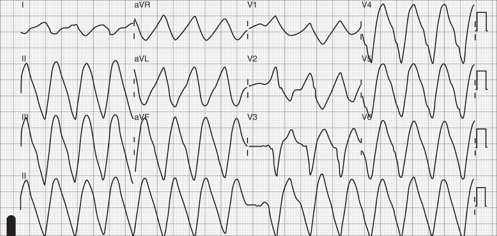
- Untreated hyperkalemia will progress to a sinuventricular rhythm or a sine wave
Left Bundle Branch Block
- LBBB as well as any LBBB configuration (ex: RV pacing) can result in ST segment elevation, usually < 5mm
- Use Sgarbossa Criteria to determine if there is a concurrent infarct
- In addition, may look for Cabrera’s sign or Chapman’s sign if infarct is suspected, though both are specific but poorly sensitive
- RBBB does not typically give ST elevation, therefore in cases of RBBB, the usual STEMI rules apply
Early Repolarization
- Normal variant often seen in young athletes
- Synonymous with J-point elevation (though not to be confused with a J-wave) i.e. elevation of the point where the QRS usually meets the isoelectric line
- Some studies suggest an increased risk of VF in these patients, though the lifetime risk remains unclear
Ventricular Hypertrophy (Left Ventricular Hypertrophy)
- LVH typically with ‘strain’ pattern: in these cases, the ST elevation should only be in V1-3, be concave-up (i.e. not a tombstone morphology), be discordant with the deep S wave, and not be elevated >2mm
Aneurysm (Ventricular Aneurysm)
- After an MI, the walls of the ventricles can become aneurysmal and manifest on the surface 12 lead as persistent ST elevation in the territory of the old infarct
- Q waves (from the previous MI) should be present in the leads with persistent ST elevation
- An echo is required for the final confirmation
- Takotsubo cardiomyopathy (broken heart syndrome) will present similarly.
Thailand (Brugada Syndrome)
- Cause by a mutation in a cardiac sodium channel (mostly SCN5A), was first described in Thailand in 1992
- May be responsible for 4-5% of all sudden cardiac deaths
- 3 described ECG types - Types 1 and 2 more commonly give ST elevation while type 3 has a “saddle back” appearance without ST elevation
- Brugada pattern can be pharmacologically induced (ex: antiarrhythmics such as sodium channel blockers), precipitated by illness or fever, or be intermittent (will commonly see an incomplete RBBB pattern)
Inflammation (Pericarditis)
- Look for diffuse ST elevation
- PR depression is typically only seen in viral pericarditis, though the teaching is that this is a classic electrocardiographic sign of pericarditis
- In acute pericarditis, there might be PR elevation and ST depression in aVR only, but this is poorly sensitive
- Consider the diagnosis of STEMI in favor of pericarditis when: there is ST depression anywhere (except for V1, aVR), ST elevation height in III>II, there is a convex/horizontal ST elevation morphology, or when there are new Q waves
- If predominantly inferior elevation, depression in aVL is very sensitive for STEMI[2]
Osborn (J) wave
- Hypothermia, usually <30 C is associated with the presence of Osborn J waves
- Positive deflections at the J point.
- Bradycardia (including AV block) and atrial fibrillation are also common in moderate and severe hypothermia
- Hypothermic patients are at risk for VF
Non-Ischemic Vasospasm
- True ST elevation, in the sense that the ST elevation pattern is that of an injury current, but has a different mechanism and a different management
- Cocaine-induced ST elevation secondary to vasospasm should be treated with benzodiazepines and nitrates as needed
- While it is possible to have a STEMI from a ruptured plaque and subsequent clot formation in a patient with cocaine toxicity, it is helpful to risk stratify patients with a suspected STEMI by age, risk-factors, etc
- It may be impossible to tell by surface ECG (and therefore without a left heart catheterization) if the ST elevation is due to cocaine toxicity or due to plaque rupture
See Also
References
- ↑ Riley RF, Newby LK, Don CW, et al. Diagnostic time course, treatment, and in-hospital outcomes for STEMI patients presenting with non-diagnostic initial ECG: A report from the Aheadache mission: lifeline program. Am Heart J. 2013; 165(1):50–56.
- ↑ Bischof JE, Worrall C, Thompson P, et al. ST depression in lead aVL differentiates inferior ST-elevation myocardial infarction from pericarditis. Am J Emerg Med. 2016; 34(2):149-154.
Authors
Max Hockstein, Neil Young, Ross Donaldson, Daniel Ostermayer