We need you! Join our contributor community and become a WikEM editor through our open and transparent promotion process.
Ultrasound: In Shock and Hypotension
From WikEM
(Redirected from RUSH protocol)
Contents
Background
- Several techniques are available for differentiating shock states
- RUSH Protocol was conceived in 2008 and looks are 3 basic aspects of physiology[1]
- The Pump
- RV:LV
- Squeeze
- Pericardial effusion
- The Tank
- The Pipes
The Protocol
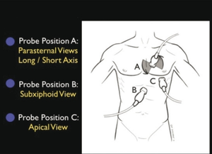
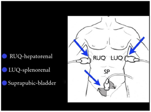
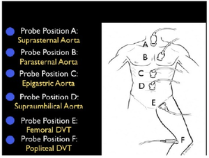
Rapid Ultrasound for Shock and Hypotension(RUSH) using the HI-MAP approach[2]
- H - Heart (parasternal and four-chamber views)
- I - Inferior Vena Cava (for volume responsiveness)
- M - Morison’s pouch (i.e., FAST exam) and views of thorax (looking for free fluid)
- A - Aortic Aneurysm (ruptured abdominal aneurysm)
- P - Pneumothorax (i.e., Tension pneumothorax)
Heart
Technique: see Cardiac ultrasound
- Pericardial Effusion
- Parasternal long
- Change in size <30% between sys and dia = poor LV function
- RV collapse
- In 4-chamber view, RV should be <60% of LV; if larger think RV failure
- Hyperdynamicity
- Walls move >90% or touch at end of systole
- May indicate hypovolemia or sepsis
- Walls move >90% or touch at end of systole
IVC
Technique: see IVC ultrasound
- Measure 2cm from RA-IVC junction
- If IVC <1.5cm and collapses on inspiration then CVP is low
- If IVC >2.5cm and noncollapsing then CVP is high
- Suggests fluid unresponsive; patient requires inotropes
Morison's
Technique: see FAST exam
- Assess for free fluid
- Morison's pouch
- Splenorenal
- Bladder
Aorta
Technique: see Aortic ultrasound
- If >5cm assume ruptured AAA until proven otherwise
Pulmonary
Technique: see Ultrasound: Lungs
- Assess for subpleural interstitial edema
- Look for multiple comet tail artifacts or "B lines"(a few, 3-4, are OK)
- If multiple found, there is interstitial edema
- Look for multiple comet tail artifacts or "B lines"(a few, 3-4, are OK)
- Assess for pneumothorax
- Scan longitudinally in anterior 2nd-3rd IC space, mid-clavicular line
- Look for lack of sliding or "beach sign"
Other
Classic Ultrasound Findings For Critically Ill Patients
| Disease | IVC | Cardiac | Lung (Phased Array) | Lung (Linear) |
|---|---|---|---|---|
| MI | ↑ | Focal WMA Mod/Poor squeeze |
NL or B-lines | Sliding |
| Tamponade | ↑ | RA collapse with filling RV collapse with filling |
NL | Sliding |
| PTX | ↑ | NL or Hyperdynamic | Lung point Consolidated lung |
Absent lung sliding |
| Sepsis | ↓ | Hyperdynamic squeeze | NL (see pneumonia) | Sliding |
| Pneumonia | NL or ↓ | Hyperdynamic squeeze | Unilateral B-lines | Sliding |
| Decompensated HF | ↑ | Mod/Poor squeeze | Bilateral B-lines | Sliding |
| PE | ↑ | RV > LV McConnell's sign |
NL or Unilateral B-lines | Sliding |
Video
See Also
References
- ↑ Weingart - http://emcrit.org/ultrasound/The%20RUSH%20Examfinal.htm
- ↑ Dina Seif. Bedside Ultrasound in Resuscitation and the Rapid Ultrasound in Shock Protocol Critical Care Research and Practice Vol 2012 ../docss/503254.pdf