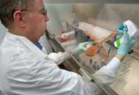
CDC scientist Steven Hurst prepares tubes for inoculation of material suspected to contain fungal organisms during the multistate meningitis outbreak.
|
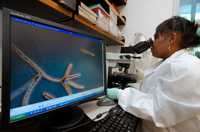
CDC scientist Carol Bolden examines microscopic slides showing Exserohilum rostratum (on screen) during the multistate meningitis outbreak.
|
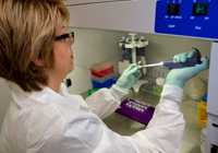
CDC scientist Christina Scheel processes spinal fluid samples for molecular testing during the multistate meningitis outbreak.
|
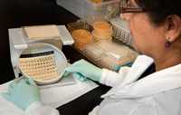
CDC scientist Naureen Iqbal reads results of antifungal drug susceptibility testing during the multistate meningitis outbreak.
|
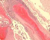
Microscopic examination of tissue from patients with clinical cases of meningitis show evidence of pus, inflammation of blood vessels, dying tissue, and blood clots.
|
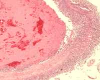
Microscopic examination of tissue from patients with clinical cases of meningitis show evidence of pus, inflammation of blood vessels, dying tissue, and blood clots.
|
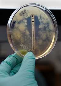
During the multistate meningitis outbreak, a plate shows results of susceptibility testing to the antifungal drug amphotericin B. The drug inhibits growth of Exserohilum in the clear area where the amphotericin drug has diffused into the medium. Exserohilum is growing elsewhere on the plate.
|
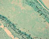
Special stains were used for microscopic examination of brain tissue from patients with clinical cases of meningitis. The black objects seen in these images is fungus.
|
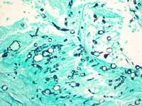
Special stains were used for microscopic examination of brain tissue from patients with clinical cases of meningitis. The black objects seen in these images is fungus.
|
 ShareCompartir


