


Immunohistochemistry (IHC)
 ShareCompartir
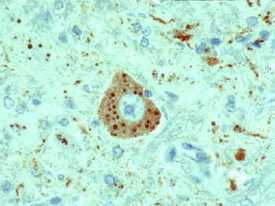
This slide shows a rabies-infected neuronal cell with intracytoplasmic inclusions. The red stain indicates areas of rabies viral antigen by using IHC or avidin-biotin complex (ABC) technique.
IHC methods for rabies detection provide sensitive and specific means to detect rabies in formalin-fixed tissues. These methods are more sensitive than histologic staining methods, such as H&E and Sellers stains. Like the dFA test, these procedures use specific antibodies to detect rabies virus inclusions. The techniques use enzyme-labeling systems that increase sensitivity. In addition, monoclonal antibodies may be used to detect rabies virus variants.
- Page last reviewed: April 22, 2011
- Page last updated: April 22, 2011
- Content source: